

Mauna Kea Technologies
- Home
- Companies
- Mauna Kea Technologies
- Applications
- Microscope System for Peripheral Nerve ...
Microscope System for Peripheral Nerve Imaging - Monitoring and Testing - Laboratory Equipment
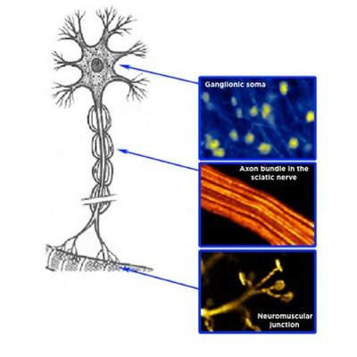
Use Cellvizio Lab to perform longitudinal studies on the peripheral nervous system.
Most popular related searches
laboratory equipment
microscopy system
laboratory testing system
microscopy
laboratory testing
monitoring laboratory
In vivo imaging of degenerating axons and neuromuscular synapses
- small skin incision is made to expose the tibial nerve and the adjacent gastrocnemius muscle
- intramuscular axons and motor nerve terminals in an innervated thy1.2-YFP16 mouse gastrocnemius muscle with an intact sciatic nerve.
- Axons in the tibial nerve of thy1.2-YFP16 mice all degenerated by 3 days after section of the sciatic nerve, in contrast to the extensive axonal protection seen in WldS mouse nerves.
- Axotomized tibial nerve axons are also preserved 3 days after sciatic nerve section in thy1.2-YFP16-WldS heterozygotes
- Intact NMJs in the gastrocnemius muscle of a WldS homozygote were observable 3 days after sciatic nerve injury but,
- No intact NMJs were ever seen in thy1.2-YFP/WldS heterozygotes at this time point.
Article reference: Axonal and neuromuscular synaptic phenotypes in WldS, SOD1G93A and ostes mutant mice identified by fiber-optic confocal microendoscopy. Molecular and Cellular Neuroscience, 10.1016/j.mcn.2009.08.002
Stay in the loop!
Select your areas of interest to receive industry updates.