

- Home
- Companies
- Creative Bioarray
- Products
Creative Bioarray products
Technology
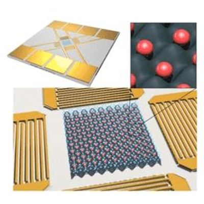
Creative-Bioarray - Patterning Cells Using Acoustic Traps
Over the past decade, remotely controlling the acoustic manipulation of cells in space within carrier matrices has emerged as a particularly promising approach for a wide range of applications, including chemical reaction control, microrobots, drug delivery, and cell and tissue engineering. Accurate control of cell positioning is a much-needed feature in tissue engineering applications, as it allows for a controlled filling of the matrix rather than relying on random seeding. Acoustic tweezers manipulate cells through the interaction of sound waves with solids, liquids, and gases, and can be used in almost any medium. Acoustic tweezers have advantages over magnetic tweezers that require magnetic particles.
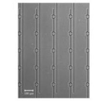
Creative-Bioarray - Patterning Cell Using Dielectrophoresis Traps
The ability to place cells in desired locations has become an increasingly important tool for controlling the cellular microenvironment. Cell patterning can be used to manipulate cell-cell interactions, thereby altering the contact area between two cell types in co-culture. Cell patterning can also be used to direct cell-matrix interactions, controlling the contact area with the extracellular matrix (ECM). Cell patterning techniques allow for a more in-depth study of the fundamental features of cells, and it has become an ideal tool for studying everything from cell behavior to molecular expression.
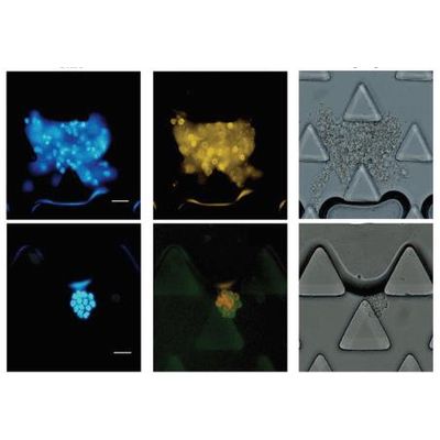
Creative-Bioarray - Patterning Cell Using Hydrodynamic Traps
In traditional cell studies, cells are often studied in larger populations, where measurements can only reflect the average of the responses of multiple cells. However, this approach can lead to misunderstandings because it masks important information about cells and their statistical properties. In contrast, single-cell analysis methods offer the possibility to extract detailed information about inherent cell-to-cell variation in large populations, providing a deeper understanding of cellular dynamics and providing high-level modeling and systems biology solutions. Quality statistics. Microfluidic separation and capture of single cells and cell clusters has shown great potential in advancing medical and biological research. The use of microfluidic devices can realize the study of intercellular interactions and signal transduction, the study of tumor cell heterogeneity and the study of drug response.

Creative Bioarray - Patterning Cell Using Magnetic Traps
Cell patterning and cell manipulation are for better control and understanding, and their application in practical biomedicine. In this regard, nanobiotechnologists have developed and implemented new methods to immobilize cells on substrates in a controlled manner, so-called cell patterning. Cell patterning and cell manipulation currently represent fundamental steps for conducting drug testing experiments as well as conducting basic research in the field of biology. Among them, magnetic manipulation refers to the manipulation of cells using permanent magnets or electromagnets. Since cells often lack paramagnetism or diamagnetism, immunomagnetic beads are used for cell surface labeling. Surface-modified magnetic beads can adhere to the cell surface through the specific binding of antibodies to antigens.
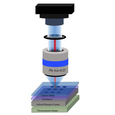
Creative-Bioarray - Patterning Cell Using Optical Traps
Cell spatial patterns and geometric constraints are considered important factors in tissue development and disease. In order to study these factors, it is necessary to provide a useful model system for cell culture. However, the ability to localize cells with specific geometries in traditional cultures remains limited, especially at the single-cell level. As a precise method, patterning techniques allow for a more in-depth study of the fundamental features of cells, and are emerging as ideal tools for studying comprehensive heterogeneity ranging from cellular behavior to molecular expression.
Creative-Bioarray - Cell Using Photolithography Traps
The ability to generate protein and cellular patterns on surfaces is important for biosensor technology, tissue engineering, and for basic research in cell biology. Certain bioassays, combinatorial screening, and biosensor fabrication all require the placement of biological ligands at well-defined locations on a substrate. Control over cellular localization is also important for cell-based screens, where repeated visits to individual cells are required to perturb them and monitor their responses. In addition, tissue engineering may also require placing cells in specific locations to create organized structures.
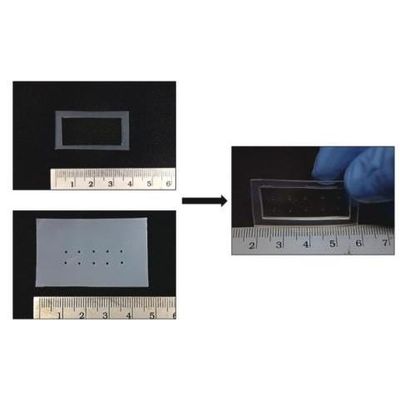
Creative-Bioarray - Cell Using Stencil Traps
Currently, cell patterning and isolation methods often utilize microfluidic systems, in which fluid forces are used to manipulate and transport cells. Inkjet-based cell "printing" and deposition methods have been shown to be effective at sorting and patterning cells at the bulk and single-cell level, but are generally low-throughput and can cause cellular stress responses. Additionally, although Trap-based methods have high throughput, but they may discriminate between specific cell morphologies or sizes associated with human disease. The microfluidic trap environment also makes it difficult to transport individual cells into isolated microenvironments for further experiments. Another disadvantage of all microfluidic patterning is that they subject cells to shear stress that can affect cell activity, function, and gene expression.
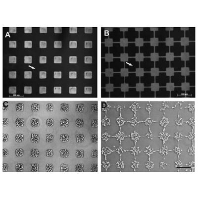
Creative-Bioarray - Cell Using Microcontact Printing Traps
The ability to pattern proteins and other biomolecules on a substrate is important for capturing the spatial complexity of the extracellular environment. Microcontact printing was initially achieved by using polydimethylsiloxane stamps to create patterns of functionalized chemicals on the surface of the material. An important part of the microcontact printing process is the topological master, from which stamps are projected; the raised and lowered areas of the master are mirrored into the stamps and define the final pattern. Typically, the master consists of a silicon wafer coated with photoresist, which is then patterned by photolithography.
Cell Analysis Kit - Cell Apoptosis Assay Kit - Caspase Assay Kit
SuperQuick - Model CSK-XA0008 - Caspase Colorimetric Assay Kit
Activation of ICE-family proteases/caspases initiates apoptosis or other cellular processes in mammalian cells. The SuperQuick Caspase-1/ICE colorimetric protease assay kit provide a simple and convenient means for assaying the activity of caspases that recognize the sequence YVAD. The Assay is based on spectrophotometric detection of the chromophore p-nitroanilide (pNA) after cleavage from the labeled substrate YVAD-pNA. Storage-20°C.
SuperQuick - Model CSK-XA0009 - Caspase Fluorometric Assay Kit
The SuperQuick® Caspase-1/ICE fluorometric assay kit provides a simple and convenient means for assaying the activity of caspases that recognize the sequence YVAD. Storage-20°C.