Delmic
5 products found

Delmic products

Cl Solutions

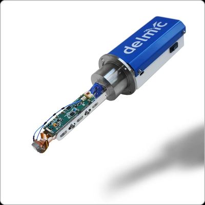
Panchromatic and RGB CL Intensity Detection

Detecting cathodoluminescence can be easy and fast with user-friendly JOLT detector. The JOLT is the youngest member of our cathodoluminescence (CL) solutions family. The JOLT is an entry-level CL-intensity detector based on state-of-the-art solid-state detectors. It comes in two variations, a panchromatic and an RGB (coloured CL) version. Adding it to your SEM can’t be easier: it is compatible with all SEM models, can be mounted on a single vacuum port and is controlled by a small software app in combination with the SEM control computer. It is a great addition for correlative imaging with other SEM-based detectors such as backscattered electron imaging and energy-dispersive x-ray imaging, capable of providing more insight into your sample’s properties. It is perfect for those who are working with and studying geological samples, semiconductors and other materials.

High-End Cathodoluminescence Intensity Detector

Harness the power of light and learn more about your material with one compact solution. SPARC Compact is a high-end cathodoluminescence intensity detector, with colour-filtering capabilities. Easily integrated in SEM, SPARC Compact is a reliable solution for providing insights into processes such as crystal growth zonation in  geological samples, and defect structures in semiconductors. This system is highly modular, which means that it can be upgraded and enhanced as your research evolves and moves forward. And just like the system, Delmic’s team is always there to support you and make sure that the high-quality results you acquire bring you one step closer to your goal.

Cryo Solutions

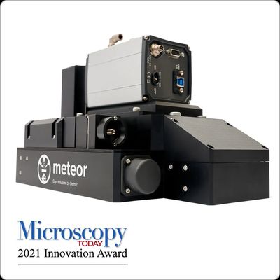
Integrated Top Down Cryo-Clem Imaging System

METEOR overcomes the challenges in the current cryogenic electron tomography (cryo-ET) and cryo correlative light and electron microscopy (cryo-CLEM) workflows by providing the capability to perform in situ fluorescence light microscopy (FLM) in your cryo focused ion beam (FIB)/ scanning electron microscope (SEM) chamber. In addition to reducing the number of transfer steps between microscopes, protecting the fragile sample from unnecessary contamination, METEOR allows you to collect fluorescence signals from the samples directly after milling. For cryo-ET you can confirm the presence of the region of interest (ROI) in the lamella to better spend your cryo-transmission electron microscope (TEM) time. For the volume cryo-CLEM workflow you can acquire better resolution FLM images throughout the depth of the sample. METEOR is highly adaptable to your current workflow and works well with your set-up, making it easy to adopt.

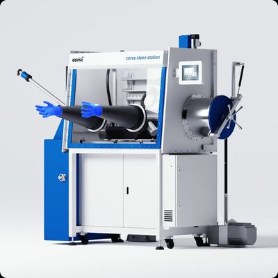
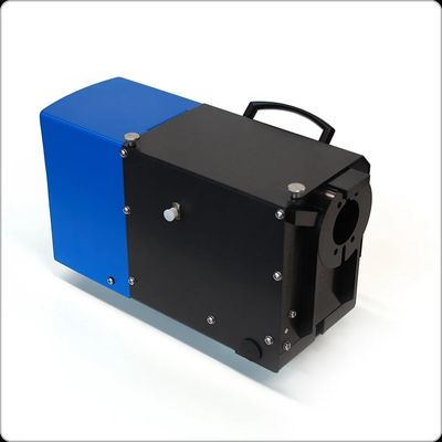
CERES Clean Station & Vitri-Lock

A common challenge in the cryo-EM workflows is to keep samples vitrified and free from ice contamination. Every scientist knows that the key to high quality data is to have good samples. Crystalline ice formed on or in the sample can obscure the region of interest and significantly lower the cryo-EM data quality. CERES Clean Station provides a clean and moisture free environment for cryo-EM users to prepare samples with ease. It is designed for high effectiveness with the comfort of the user in mind. CERES Vitri-Lock uniquely enables your cryo-sample transfer to take place in a high vacuum, anhydrous environment, to ensure your samples stay uncontaminated during the transfer between the Clean Station and the cryo-FIB/SEM.

Fast Imaging

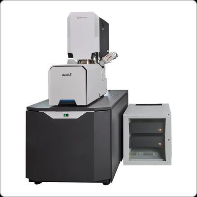
Ultra-Fast Automated Multibeam Electron Microscope

Are you looking to image 3D EM volumes of unprecedented scale? Meet FAST-EM, an ultra-fast automated multibeam electron microscope (EM), designed together with ThermoFisher, Technolution and TU Delft to make complex EM  workflows simple and efficient. FAST-EM can image biological thin samples at unprecedented speeds, and with a level of automation that enables large scale imaging without the need for constant supervision. Delivering powerful insights while keeping the workflows simple, this system allows you to shift the focus from microscope operation to data analysis. FAST-EM can be used to explore cell architecture, the interaction of neuronal circuits, and the analysis of any biological material in life sciences. It is extremely beneficial for large volume 3D imaging, large scale 2D imaging and, in general, as a tool that can significantly speed up daily microscopy facility work.