

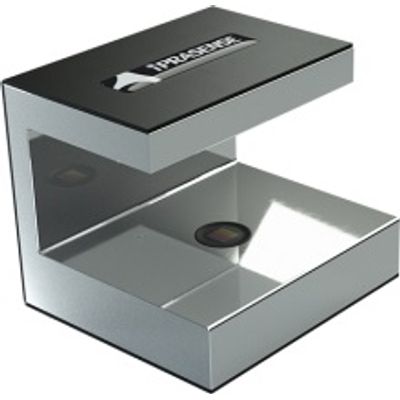
Flotek iPRASENSE Cytonote - Model 1W -Cells and Real-Time Analyzer
Time-lapse images of cells and real-time analysis from inside your incubator; The Cytonote is designed to be compatible with every standard lab dishes, T-Flask or slides. Select your favorite container from your favorite manufacter and place it on the Cytonote. If you want, modify the delay between 2 acquisition (default: 20min) and your culture is ready to be recorded over long period of time. As explained, no need to adjust focus, brightness or any other parameters, our algorithm will reconstruct cells wherever cells are located... from a few micrometers to some millimeters! This caracteristic mades Cytonote a extermely robust device not fearing temperature variation, cells descent inside matrigel, or any other parameter which can affect traditional focus adjustment.
Our innovative instruments open new perspectives into Live Cell imaging and cell kinetic analyses. IPRASENSE’s label-free time-lapse Imaging Technology offers a versatile solution for monitoring cell culture inside your incubator. The unmatched extra large field of view and the insensitivity to focus provide a robust real-time analysis of your adherent cells in any Petri dishes, T-Flask, slides or microchips.
The CYTONOTE 1W product range simplifies live cell imaging technique and transforms the complex and expensive microscope into a cost-effective solution.
The CYTONOTE 1W is a the most simple live cell-imaging system designed for recording cell movies and analysing a variety of cell culture from inside the incubator. The innovative and patented « lensless imaging » technology pushes the boundaries of microscopy with its super wide Field of View and its capability to capture and analyze precisely several thousands of cells without any focus and brightness settings.
The image analysis and results from the Cytonote are performed from the HORUS dedicated software. HORUS is application oriented, it provides automatic cell count, quantitative confluence determination, cell size or cell tacking. Full field images (30 mm2 ) of the samples are stored and can be accessed and zoomed at any time. lt is designed to monitor up to 6 Cytonote simultaneously for 6 parallel or independent cell cultures.
- CELL MIGRATION CHEMOTAXIS, WOUND HEALING ON HIGH STATISTICAL NUMBER OF CELLS AND VERY WIDE AREA
- CELL PROLIFERATION THROUGH CELL COUNT AND QUANTITATIVE CONFLUENCE DETERMINATION
- ANGIOGENESIS THE VERY WIDE AREA ALLOWS TO OBSERVE THE FULL ANGIOGENESIS PROCESS WITH HIGH LEVEL OF DETAILS
Cells: Eucaryotic cells : adherent monolayer, suspension cell at bottom of culture ware or in micro-slides, 3D spheroids
Media: Liquid or semi-solid (collagen)
Culture vessels: Standard plastic petri dish, culture flasks, multiwell plates, max height 55 mm
Resolution: 1 micron
Field of view: 29,4 mm2
Working distance: 0 to 5 mm
Image rate: 1,5 min
Light source: LED
Sensor: CMOS 10 Mplx
Dimensions: 12 x 11 x 10 cm
Weight: 1 kg
Power supply: USB