

- Home
- Companies
- Maltron International – Leading ...
- Products
- Maltron - Model Sheffield MK 3.5 - ...
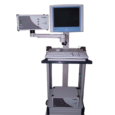
Maltron - Model Sheffield MK 3.5 -Electrical Impedance Tomography (EIT) Device
The Maltron Sheffield MK 3.5 – Electrical Impedence Tomography (EIT) device – is a state of art Impedance Imaging research and clinical tool that has applications in Pulmonary, Gastric and Body Segment monitoring, as well as Tissue characterisation.
Over 20 years of research and development in the field of Electrical Impedance Tomography has provided a state of art tool for a number of significant medical applications, as a safer and more cost effective method of assessing patients.
The Maltron Sheffield Mk 3.5 applies a set of electrodes around the area under investigation and then makes a series of impedance measurements. These measurements are then used to produce images.
Many of our designs have already been implemented, and the MK1 and SAPT systems have been especially popular for medical research groups the world over – making Maltron Sheffield the largest manufacturer of EIT systems in the world.
The latest MK3.5 was designed primarily for ease of applications and its use on neonates.
Over 2 decades of history of development which has resulted in the following products being implemented:
- 16 Electrodes – 50KHZ data collection system
- Miniature version of MK1, for use with astronauts and flown on the Russian MIR programme
- 16 Electrodes – 20KHZ.
- 16 Electrodes – Operating at 8 frequencies from 9.6KHZ TO 1.2MHZ
- 64 Electrodes – Operating from 9.6KHZ TO 1.2MHZ
- 8 Electrodes – 2KHZ to 1.6MHZ at 30 frequencies.
Bone is a poor conductor, whereas muscle is a relatively good conductor.
Biological tissues conduct electricity as they contain ions, which act as charge carriers. Some tissues conduct electricity better than others because they contain more ions that are free to carry charge.
The MK 3.5 offers several advantages over other body imagining and monitoring methods:
- Low Cost
- No known hazards
- Long term monitoring of Physiological function is possible
- Rapid data collection so that changes in function can be measured
- Can use spectral measurements to make tissue characterisations
- Technique : Impedance Analyzer Tomography
- Frequency : Multi-Frequency 2khz – 1620 khz
- Channels : 8
- Impedance Range : 1-100 Ohms
- Data Collection Speed : 25 frames/sec
- Accuracy No. Profile Images : c.1.0% 12bit ADC8
- Demodulation : Digital
- Outputs : High Speed Serial to PC isolated to IEC601
- Computer Interface : High Speed Serial PCI card
- Patient leads : 8 Triaxial
- Data point images : 64
- Drive current : 200uAp-p at each frequency conforming to IE601
- Software : Matlab
- AT Environment : +10oc to 40oc
- Relative Humidity : 30% to 75% non-condensing
- Atmospheric Pressure : 700KPa to 1060KPa
- Service : No serviceable parts other than periodic calibration
- Guarantee : 12 months parts & labour (excluding disposables)