

- Home
- Companies
- Evident Scientific
- Products
- Evident - Model SLIDEVIEW VS200 - Slide ...
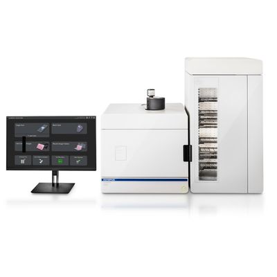
Evident - Model SLIDEVIEW VS200 -Slide Scanner
Perform applications from high-end research to specialized digital pathology with our most advanced slide scanner. The SLIDEVIEW™ VS200 system is designed to capture a wide variety of samples and stains with multiple magnifications, flexible slide sizes, and five versatile imaging modes. Outstanding image quality of whole slides empowers your lab to unlock the full potential of your samples.
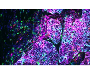
The SLIDEVIEW VS200 system offers high-resolution and high-throughput slide scanning for advanced research applications, such as neuroscience, cancer and stem cell research, and spatial biology. With the flexibility of five imaging modes, multiplexing capabilities, and multiple magnifications up to 100X, the system delivers outstanding image quality for quantitative analysis.
SLIDEVIEW VS200 for Digital Pathology
As a flexible yet intuitive scanner, the SLIDEVIEW VS200 system enables your team to digitize a wide range of sample types with minimal training. The VS200 system helps you achieve a fully digital workflow for pathology with versatile slide scanning, outstanding image quality, and flexible integration options. Get the evidence you trust for standard and specialty samples to make breakthrough decisions with confidence.
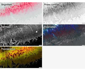
Discover more details in your samples with five imaging modes—brightfield, polarization, fluorescence, darkfield, and phase contrast in one system—and the ability to combine multiple techniques in a single scan.
Supporting up to six high-performance X Line™ objectives and featuring True Color LED illumination and color-corrected camera profiles, the SLIDEVIEW VS200 system delivers clarity in whole slide images.
The True Color LED for transmitted illumination has the same spectral characteristics and power as a halogen lamp, so purple, cyan, and pink stains are correctly represented, imaged, and rendered.
- Enhanced Z-focus with linear encoders offers higher readjustment accuracy.
- Automatic sample detection helps define the scan area for faster imaging, with the option to manually adjust if needed.
- Autofocus improves the automated stitching function.
- Design minimizes vibrations for better imaging stability.
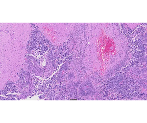
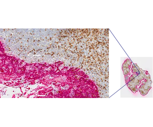
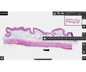
Tonsil CD3 (rm), ImmPRESS Reagent (HRP) Anti-Mouse IgG Immpact DAB (brown), AE1/AE3(m) ImmPRESS (AP) (HRP) Anti-Rabbit IgG Immpact Vector Red (red). Counterstained with Hematoxylin QS (blue). Image data courtesy of Vector Labs.
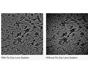
The fluorescence illuminator’s fly-eye lens uniformly distributes light across the entire field of view for bright, evenly illuminated images.
Accelerate workflows with a high-capacity loader that holds up to 35 sample trays, accommodating up to 210 slides (26 × 76 mm / 1 × 3 in.). Robotic automation ensures fast, precise loading and unloading, coordinating with the focus and scan unit for rapid image acquisition, while an integrated barcode reader automatically captures and records slide information.
- Identical settings mode: Automatically assigns scan settings to all the slides.
- Individual settings mode: Change specific settings for each slide or all slides in a single tray.
- Flexible batch scan mode: Assign different observation methods, such as FL, BF, POL, DF and PH, for each slide contained in the batch.
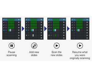
- Priority scan function: Interrupt an ongoing batch to scan a priority slide before resuming.
- Hot-swap: Replace scanned trays without pausing the batch process, minimizing downtime.
- Efficient and flexible: The software is easy to set up and operate. You can upload your trained neural network and open it with the click of a button.
- Reduce the volume of acquired data: TruAI technology and selective scanning prevents the system from acquiring large amounts of data from areas you don’t need. This helps optimize data management, including storage, uploading, and image sharing.
- Set it and forget it: TruAI technology is designed to support accurate region detection based on trained parameters, reducing the need to supervise the scanning. This enables you to use your time for other tasks and do scanning runs overnight or over the weekend.
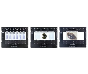
- Mixed-batch scanning: An easy-to-use slide tray enables you to mix 26 × 76 mm (1 × 3 in.), 52 × 76 mm (2 × 3 in.), 76 × 102 mm (3 × 4 in.), and 102 × 127 mm (4 × 5 in.) slides on the same batch scan.
- Automated slide detection: An RFID system detects different formats and scans them in the same batch, saving time and maximizing throughput.
- Centralized image storage: Conveniently manage your images with the optional Net Image Server (NIS) SQL database.
- Secure web-based sharing: Easily store and share images online to collaborate with colleagues, research teams, or remote consultants.
- Access control: Control access to your data with individual access rights.
- Multiple file formats: Scan and export images in VSI, OME-TIFF, and DICOM formats.
- LIS integration: Query the laboratory information system (LIS) for additional metadata and save scans in DICOM format for direct upload to an image management system.
- Automated workflows: Enable automatic uploads to the NIS SQL database for streamlined data management.
- VSI format for advanced microscopy: Developed by Evident, the VSI file format meets the specialized needs of microscopy imaging. Its growing adoption by various projects and companies ensures long-term compatibility and support.
- View virtual slides on any Windows PC: locally or over a network.
- Remote access: Open and analyze images saved on the NIS via the internet.
The integrated barcode reader automatically captures and records slide information, simplifying slide tracking and data entry.
- Supports a wide range of 1D and 2D formats for broad compatibility.
- Data can be configured as a file or folder name, simplifying the organization and management of large slide volumes.