

Guava Muse - Cell Analyzer
Sophisticated cell analysis doesn’t have to be complicated or costly. The Guava Muse Cell Analyzer is an easy to use, simple, compact, robust instrument for your cell analysis needs. With the Guava Muse Cell Analyzer, you can now achieve highly quantitative results at a fraction of the price, effort, and time. The Muse Cell Analyzer packs 3-parameter analysis into a compact, easy to use benchtop device, making flow cytometry accessible to anyone, anytime. A user-friendly touchscreen interface, intuitive cell analysis software, and optimized assays work to simplify your research.
- Quantitative data at the single-cell level
- Simple and effortless operation
- Intuitive cell analysis software and touchscreen user interface
- Rapid setup and analysis
- Optimized Muse assays
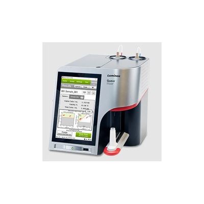
- Compact size: footprint of only 8 in x 10 in (20 cm x 25 cm)
- Flow cytometry technology in an affordable instrument
The Muse Instrument has an integrated touchscreen and software for data acquisition and analysis using optimized Muse Assays. Its microcapillary flow cell is engineered for acquisition of both suspension and adherent cells 2-60 microns in diameter.
Sample collection, acquisition, and analysis of results is achieved in just a few steps. The user interface is designed to be intuitive, so you spend less time with experimental setup and analysis for the critical cell health assays you need most.
The system uses a microcapillary and miniaturized optics, which occupy one-tenth the space of a typical cytometer. This means that the instrument occupies only an 8 in x 10 in (20 cm x 25 cm) footprint. A green diode laser is used for excitation, and a uniquely designed series of retro-reflective lenses provide maximum light capture and sensitivity.
Muse Software is designed to be simple enough for any user to follow setup instructions to achieve sample acquisition, on the first use. Assay-specific software modules include sample graphs to make population gating setup foolproof. Results are displayed in both graphical and statistical formats specific to each application, making analysis unambiguous.
The figure below shows the data output for the Muse Count & Viability Assay. Healthy Jurkat cells were mixed with heat-killed Jurkat cells and stained with Muse Count & Viability Reagent, and then analyzed on the Muse Cell Analyzer. Data output include summary data (not shown) and optional dot plots (shown here). Reported statistics include viable cells/mL, % viability, and the total cells/mL. The left hand dotplot shows viability vs. cell size; the right hand plot shows viability vs. nucleated cells.
Multiple adherent and suspension cell types were harvested, diluted, and counted. Cell counts from all three methods were averaged to obtain “theoretical cell concentration”. Each point represents the average cell concentration of 3 replicates, and each data series was fit with linear regression. Muse cell analysis data was correlated with the theoretical concentration with slope closest to 1, indicating superior accuracy.
The concentration measurements for serial dilutions of five representative cell lines are shown, and include both adherent and suspension cells. Each point represents the average of three replicates. Expected cell concentrations were calculated by measuring the stock sample concentration with the Muse Cell Analyzer, then dividing by the dilution factor to obtain theoretical concentrations of diluted samples. Data show the comparison of observed vs. expected cell concentrations.