

- Home
- Companies
- Micro Photonics Inc.
- Products
- Model SkyScan 1273 - High-Energy ...
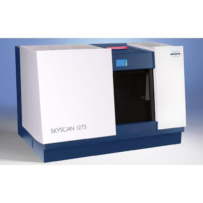
Model SkyScan 1273 -High-Energy Desktop Micro-CT
DESKTOP YET LARGE FORMAT
The SkyScan 1273 High-Capacity Micro-CT provides the ability to image large and/or dense objects that typically require larger, more expensive floor standing models, but in the space saving format of a benchtop model. The SkyScan 1273 plugs into a standard 110V wall outlet, does not require water-cooling, or compressed air making installation easy in almost any laboratory setting.
HIGH CAPACITY
The SkyScan 1273 has one of the largest chamber sizes on the market for desktop micro-CTs. The specialized rotation stage allows objects up to 25kg to be mounted without any loss in rotational accuracy.
LARGE FIELD OF VIEW
The large field-of-view CMOS detector is combined with the innovative SkyScan OFFSET scanning mode—a physical horizontal shifting of the detector to widen the field of view and increase the pixel array, without having to increase the working distance.
HIGH ENERGY
The SkyScan 1273 offers a high energy X-ray source for a desktop, with flexible modes to image very fine structures at high resolution or scale up with more power for larger denser objects.
HELICAL SCAN MODE
In addition to the standard circular scan mode, helical scanning and reconstruction can be select to optimize image acquisition and minimize reconstruction artifacts on metal objects like implants and other manufactured parts.
HART MODE
High Aspect Ratio Tomography (HART) allows for adaptive rotational steps for faster scanning of high aspect dimensional samples.
—–
IN SITU STAGES
Each system comes pre-wired to accept specially designed in situ stages for dynamic testing. The Materials Testing Stage (MTS) allows for in situ compression or tension testing of materials. Heating and Cooling Stages are ideal for material characterization for thermal effects.
SOFTWARE
The SkyScan 3D Software Suite is supplied as an unlimited site license so each user can have a copy; ensuring workflow from multiple groups is easily managed. Segmented software programs means post acquisition reconstruction, analysis, and modeling can be done on different computers, freeing the system for scanning. Image file outputs are standardized to TIF, BMP, or JPG formats for easy viewing, transferring, or copying.
- X-ray Source
40-130 kV
max. 39 W
< 5 µm spot size at 4 W - X-ray Detector
6-megapixel (3072 x 1944)
Active pixel CMOS flat-panel detector - Maximum Object Size
300 mm diameter
500 mm length
20 kg - Maximum Field of View (FOV)
250 mm height
250 mm diameter - Optional Stages – Micro positioning, cooling, heating, compression/tension
- Radiation Safety – <1 µSv/h at 10 cm from the instrument surface
- Dimensions
1250 mm (W) x 815 mm (D) x 820 mm (H)
450 kg - Power Supply
100-240 VAC, 50-60 Hz, 3 A max