

Lumencor - Model TARGA -Imaging System
The TARGA high-throughput imaging system, a dedicated assay-specific scanner, seamlessly combines the key components necessary for best data quality and speed: a high-performance solid state light engine, precision motion control, a sCMOS camera and a dedicated computer. TARGA is a highly integrated platform for efficient, fast data collection and analysis.
TARGA incorporates Lumencor’s best-in-class solid-state illumination technology. Discrete, electronically controlled light sources allow inter-channel balancing of fluorescence signals and rapid wavelength switching. TARGA utilizes between two and eight epifluorescence excitation colors, moving sample stage with stationary objective. Bright field, color-selective fluorescence, white light transmissive detection and birefringence are possible. The intrinsic stability of the solid-state lighting can be further enhanced by feedback-controlled light metering when precise quantitation is required. Light dosage can be known, routinely monitored and is stable. The objective mount is a motorized with interchange possible for two objectives that use infrared reflection-based and/or image-based, including predictive, focus to autofocus.
TARGA’s operation is controlled by an onboard server with ethernet connectivity to a client. It comes Micromanager compatible with API available for Java/C++/MATLAB/Python available. With front loading sample exchange, assays can be run autonomously, with local data storage via the onboard computer, or under client supervision, with ethernet data transfer to remote storage, and analyzed with customized image analysis for specific assays such as real-time cell counting. Multiple TARGA imaging systems at one or several locations can be calibrated for inter-instrument performance, networked, and coordinated from any client, enabling a distributed and scalable approach to high-throughput analysis. TARGA’s integration at both instrument level and as a networked appliance offers a new approach to the increasingly large-scale screens required to gain systematic insights into biological processes.
- Sample Scanner Configuration 96-, 384-, 1536-well microplate or four 75 mm x 25 mm microscope slides
- Scanner Stage Motion Resolution X=88 nm, Y=88 nm, Z=5 nm
- Standard Objectives 10X/0.45 NA air, 40X/0.95 NA air (5X/0.25 NA air, 20X/0.80 NA air & others available).
- High-resolution Objective Various magnification and NA objectives, details upon request
- Excitation Sources 5 solid state stabilized sources, with excitation filters covering 350 to 750 nm.
- Fluorescence Detection Quad-band dichroic + 4 single band emitters in fast filter wheel.
- Camera Major makes and model options, Large sensor compatible
- Focus Flatness <200 nm focal separation (center to corner).
- Illumination Flatness <30% (center to corner).
- Pixel Shift <3 pixels shift between fluorescence channels, <1 pixel shift with SW correction.
- Color Switching Time 50 msec is the minimum time to change colors and collect an image.
- Scan Time (4-color detection) 96 wells in 1.5 minutes; 384 wells in 5.1 minutes; 1536 wells in 17.8 minutes.
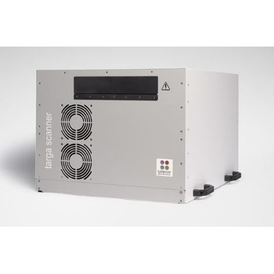
- Dimensions 20in x 23in x 16in (51cm x 59cm x 40cm)
- Weight <100 lbs (<45 kg)