- Home
- Companies
- Nippon Genetics Europe
- Products
- Nippon Midori Green Advance - Model ...
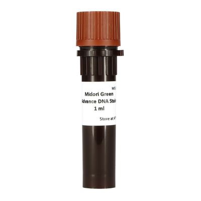
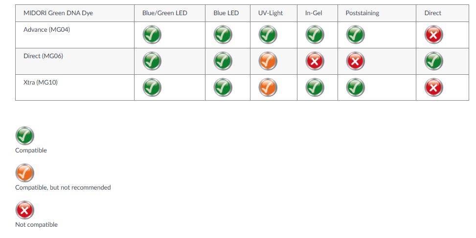
Nippon Midori Green Advance - Model MG04 -Nucleic Acid Stain Ethidium Bromide
Safe DNA/RNA stain optimal for UV-light. MIDORI Green Advance is a safe alternative to the traditional nucleic acid stain ethidium bromide. It is a non-carcinogenic and less mutagenic dye for detecting dsDNA, ssDNA and RNA in agarose gels with a very high sensitivity. MIDORI Green Advance can utilize with UV light or with our innovative Blue/Green LED technology.
- Perfect in-gel staining or poststaining of DNA/RNA in agarose gels
- No toxicity, non-carcinogenic
- Safe alternative to ethidium bromide
- High fluorescence
- Optimal for UV-light
- 1 mL staining up to 25 liters of agarose
Safe alternative to ethidium bromide
MIDORI Green Advance is a non-carcinogenic dye. Optimised for a brighter signal when excited by UV-light or Blue/Green light. It has the advantages, such as being non-carcinogenic and having an excellent signal-to-noise ratio.
MIDORI Green Advance shows a very high sensitivity even for small DNA fragments. The dilution factor of MIDORI Green Advance can be as high as 1:25000. Hence, 4-6 μL are enough for the staining of a 100 mL agarose gel, resulting in ~17 to 25 liters of stained agarose gels.

It is essential for a good replacement of the mutagenic DNA stain ethidium Bromide to deliver strong signals. MIDORI Green Advance delivers signals with a comparable intensity. Nonetheless, the safety of the user must not be compromised. Hence, several tests were performed with MIDORI Green Advance and according to those tests, MIDORIGreen Advance is safe.
- Ames-Test
- Acute Oral Toxicity Test
- Mouse Bone Marrow Micronucleus Test
- Chromosome Aberration Test
- Latex and Nitrile Gloves Penetration


- Prepare 100 mL of agarose gel solution (concentration from 0.8-3.0%) and heat until the solution is completely clear and no small floating particles are visible.
- Add 4-6 µL of MIDORI Green Advance DNA Stain to the gel solution and mix it gently. Cool the gel to 50- 60ºC and cast the gel, into the gel tray. When the gel is solid, load the samples and perform electrophoresis.
- Detect the bands using a UV or LED illuminator. Yellow or green gelatin- or cellophane filters should be used for photography.
- Data kindly provided by:Advance DNA Stain poststaining solution may be used 2-3 times.
- Staining solution that is going to be reused should be preferably stored at room temperature in the dark.
- For
- Optimal staining time (5-60 minutes) and the amount of the stain may depend on the thickness of the gel.
NOTICE: Usage is not recommended with SDS containing loading buffers because of band appearance caused by stain and SDS interaction!