

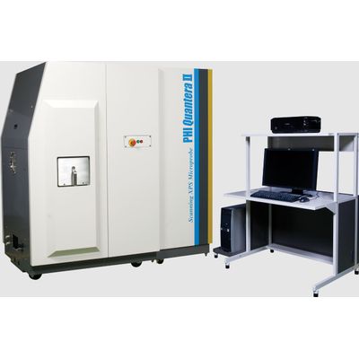
PHI Quantera - Model II -Scanning XPS Microprobe
The core technology of the Quantera II is PHI’s patented, monochromatic, micro-focused, scanning x-ray source which provides excellent large area and superior micro-area spectroscopy performance. Spectroscopy, depth profiling, and imaging can all be performed over the full range of x-ray beam sizes including the minimum x-ray beam size of less than 7.5 µm. In addition to superior XPS performance characteristics the Quantera II provides two in situ sample parking stations which enables the automated analysis of all three sample platens in a single user defined analysis queue.
- The unique scanning X-ray microprobe allows SEM like navigation with point-and-click control
- X-ray induced secondary electron imaging (SXI) provides perfect correlation between imaged areas and spectroscopy
- Multiple ion gun options (monatomic Ar, C60, argon cluster GCIB) for a variety of organic, inorganic, and mixed materials
- Full 5-axis stage functionality including rotation/tilt and heating/cooling during sputtering
- Multipoint profiling within a single sputter crater for on/off defect analysis and precious samples
- Adjustable solid collection angle for improved angular resolution for Angle Resolved analysis with advanced software for high-throughput film structure analysis
- Highest small area sensitivity on the market
- <10 microns microprobe size in x and y
- Image registration for unattended automated micro-area analysis
- Heating and Cooling in situ
- Electrochemical (biasing, polarization studies) experiments
- Glove Box Adapter
Micro-Focused Scanning X-ray Source
UNIQUE TECHNOLOGY
- Micro-focused, raster scanned x-ray beam
- X-ray beam induced secondary electron imaging
- XPS images with spectra at each pixel for retrospective chemical analysis
- Point or multi-point spectroscopy
- Point or multi-point thin film analysis
Micro Area Spectroscopy
UNIQUE TECHNOLOGY
- Micro-focused raster scanned x-ray beam
- Minimum beam size < 7.5 µm in diameter
- Confidently locate small features for analysis using x-ray beam induced secondary electron images
- Highest small area XPS sensitivity
-
X-ray beam induced secondary electron image (SXI)
- Spectra from selected areas obtained using a 20 µm diameter x-ray beam show Cu on the surface of the metal pads and SiO_2_ off of the pads.
Thin Film Analysis
INORGANIC THIN FILM ANALYSIS
- 0-5 kV floating column ion gun
- Low voltage depth profiling for ultra-thin films
- Compucentric Zalar rotation
- Effective dual beam charge neutralization Bend in column to stop neutrals
- Micro-area depth profiling
- Multi-point depth profiling
2 keV sputter depth profile of the surface species on a solder ball used for semiconductor packaging.
- Optional Ar2500+ and C60 cluster source ion guns
- Mass filtered mass ion beam
- Sputters many polymer and organic materials without damaging their chemistry
10 keV C_60_ depth profile of a 50/50 rapamycin and PLGA film showing segregation of the rapamycin to the surface of the coating.
- Robust Auto-Z sample alignment
- Turnkey dual beam charge neutralization
- Move without concern from insulator to conductor in auto analysis sequences
- No special sample mounting or masking