

- Home
- Companies
- Thermo Fisher Scientific, LIMS & ...
- Products
- Thermo Scientific Tundra - Model ...
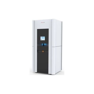
Thermo Scientific Tundra - Model Cryo-TEM -Transmission Electron Microscope
Cryo-TEM that is cost effective and easy to use, bringing cryo-electron microscopy to every biochemistry laboratory. The Thermo Scientific Tundra Cryo-Transmission Electron Microscope (Cryo-TEM) is a dedicated structure-determination solution designed to bring single particle analysis to every biochemistry laboratory. It is easier to use than typical cryo-TEM instruments, fits into a standard lab space, and matches grant mechanisms and funding opportunities globally. The Tundra Cryo-TEM is a powerful tool that can help answer your most challenging research questions, offering structural determination at biologically relevant resolutions.
Structural information at biologically relevant resolution
The optics of the Tundra Cryo-TEM are specifically designed so that you can answer fundamental questions on biology and human disease with data at biologically relevant resolutions. Our first and only microscope operating at 100 kV, the Tundra Cryo-TEM can generate high-contrast biological images at medium throughput with its high-performance, fraction-enabled Thermo Scientific Ceta Camera.
This special combination of optics and detector technology enables the Tundra Cryo-TEM to solve protein structures to biologically relevant resolution at medium throughput.
Space efficient and cost effective
The new hardware architecture of the Tundra Cryo-TEM has been purposefully designed with a smaller footprint and an easier access path without sacrificing performance. In many cases, this allows you to avoid the additional investment and unwanted downtime that comes with modification of your existing laboratory infrastructure (or even the need for a new, purposely built lab) to accommodate the instrument.
Additionally, a wide range of funding and instrumentation grants were considered when designing the Tundra Cryo-TEM so that its price falls within the reach of most instrumentation grants.
Easy, iterative sample-viability determination and biochemistry optimization
Electron microscopy is a straightforward method for the quality assessment of purified biological specimens at the microscopic scale. The Tundra Cryo-TEM can visualize the impact of biochemical adjustments to samples faster than other technologies, since you do not need to go through the lengthy process of crystals growth.
Each iteration is extremely efficient due to the instrument’s sample loading technology. It only takes a few minutes to exchange samples in and out of the microscope, allowing you to optimize sample conditions quickly. Notably, this technology is designed so that even new users are capable of doing this procedure without extensive training. The instantaneous feedback of the Tundra Cryo-TEM significantly shortens the time required for biochemical sample optimization.
Unique AI algorithms
The Tundra Cryo-TEM comes with a complete suite of automation software for efficient optimization of your sample`s biochemistry as well as data collection for structural determination. This includes user-friendly single particle analysis (SPA) data acquisition software, Thermo Scientific EPU 2 Software, guided day-to-day operation, a traffic-light UI element that indicates the microscope’s status, and pre-defined templates for typical use cases that allow you to begin collecting high-resolution data with only a few clicks.
Additionally, the Tundra Cryo-TEM is the debut of smart EPU software, an AI-enabled software solution capable of analyzing intermediate results, providing instant feedback, and steering data collection on the fly. Our AI algorithms are based on years of cryo-EM knowledge, replace decisions that experts need to make upfront, and ensure that your instrument is working at optimal conditions, allowing you to focus on the science rather than on fine-tuning the microscope.
3D structures answer your biological questions
One of the primary challenges of investigating dynamic biological processes, and how they fail in disease, is the complexity inherent to biological machinery. Large and/or dynamic protein systems present a unique challenge to traditional methods of scientific inquiry, which study these systems either indirectly or in isolation. Fortunately, cryo-electron microscopy (cryo-EM) single particle analysis (SPA) has emerged as a well-suited approach for the direct determination of native function and dynamics in complex biological systems.
Single particle analysis can validate your biochemistry work by showing the molecular details that underlie the interactions between proteins, small molecules, and post-translational modifications in large and dynamic protein systems at near native conditions. These molecular details confirm the mechanism of action by which complex biological systems (e.g. membrane proteins, protein complexes, and macromolecular machines such as viruses, ribosomes, and proteasomes) contribute to human health and disease.
For example, the human GABAA (gamma-aminobutyric acid type A) receptor is a small membrane protein and ligand-gated chloride-ion channel that mediates inhibitory neurotransmission. GABAA receptors are important therapeutic targets as their various conformations affect a variety of important signaling pathways. Due to GABAA`s conformational flexibility, traditional methods have been unable to reveal its molecular mechanism of action. The ability of cryo-EM to image large, biologically complex structures has, meanwhile, allowed researchers to see the molecular details that underpin the allosteric modulation of this important receptor.
With the Tundra Cryo-TEM, more scientists can have access to this type of information for their complex biological systems. Our applications scientists have used the Tundra Cryo-TEM to determine a 3D structure of GABAA to ~4-5 Å resolution. At this resolution, molecular details such as individual alpha helices and beta sheets are clearly visible, molecular structures can be docked, and molecular interactions modeled. Additionally, the Tundra Cryo-TEM was able to achieve 3 Å resolution for the rigid benchmark protein apoferritin. At this resolution, the protein backbone can be traced and major amino acid side chains are clearly visible, making de novo model building possible. Data at these resolutions helps you understand how proteins function, how to modify genes, and how to design drugs accordingly.
- Technical highlights of the Tundra Cryo-TEM
- 3.5 Å apoferritin structure determination in 24-72 hours
- High-brightness X-FEG (extreme field emission gun)
- Fixed accelerating voltage of 100 kV
- Semi-automated sample loading
- Cryo-preparation station allowing contamination-free sample exchange
- Sample transfer device for transferring single AutoGrids to the microscope
- Fixed cryo-box that keeps samples contamination free for up to 72 hours
- Computerized 4-axis specimen cryo-stage with ±15° alpha tilt
- High-performance Thermo Scientific Ceta Camera with dose fractionation, optimized for low-dose imaging
- EPU 2 Software for automated single particle analysis screening and data acquisition
- Smart EPU software: an AI-enabled software solution that provides feedback into data collection. Includes an open application programming interface
Floor-plan and installation requirements
- Environmental temperature: 18–23°C
- Temperature stability: 1°C per 24 hours
- The enclosure can handle any temperature variation time within this bandwidth
- Relative humidity: <80 %
- Minimum room dimensions: 4.00 x 3.80 m (13.1 x 12.5 ft)
- Ceiling height: 2.74 m (8.99 ft)
- Door height: 2.30 m (7.55 ft)
- Door width: 1.00 m (3.28 ft)
- Weight distribution maximum: 700 kg/m2
- Double earth connection
- Frequency: 50 or 60 Hz (±3%)
- Compressed air supply with a pressure range of 5–7 bar
- Sulfur hexafluoride (SF6) gas in properly ventilated room
- LAN connection for Thermo Scientific RAPID Service (Remote Access Program for Interactive Diagnosis)