
- Home
- Companies
- Nirmidas Biotech, Inc.
- Products
- DeepVision - Model ...
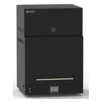
DeepVision - Model VIS/NIR-I/NIR-II/SWIR -VIS/NIR-I/NIR-II/SWIR Small Animal Fluorescence Imaging System
DeepVision Imaging Instrument for Novel NIR-I and NIR-II Dyes and Probes. An introduction video on DeepVision™ Imaging System and its Technology. In vivo fluorescence imaging of small animals has been done typically in the NIR-I (800-900 nm) range, suffering from shallow imaging depth and high background due to light scattering and tissue autofluorescence.
NIR-II/SWIR imaging is a breakthrough development detecting fluorescence/luminescence in the 1000-1700 nm range to suppress these effects, affording single cell resolution at down to ~ 3 mm depth and useful feature resolution up to ~ 1 cm depth. Combined with ultra-bright NIR-II probes (molecules, quantum dots and rare-earth nanoparticles licensed from Stanford University) from Nirmidas Biotech and a new camera technology (10X shorter exposure time and lower noise than older brands/makers), DeepVision™ affords high performance non-invasive imaging of vasculatures, tumors, intact mouse brain, lymphatic vessels/lymph nodes and molecular imaging using antibody conjugated probes. It is a new generation of NIR imaging instrument empowering researchers to interrogate cardiovascular, cancer, brain and immune disease models. NIR-II/SWIR imaging is generating thousands of publications in recent years and is ideal for deep-tissue animal imaging in vivo.
In addition to CW imaging mode, a unique added feature of DeepVision™ is its fluorescence lifetime imaging mode. By setting a delay time of detection, the emission generated by a 975 nm excitation pulse from rare-earth nanoparticles (lifetime ~ 10 milliseconds) versus the emission from quantum dots (lifetime in micro-second range) can be distinguished despite their overlapping emission wavelengths. Such life-time imaging detects zero background. This scheme can also be used for multiplexed molecular imaging in the least scattering 1500-1700 nm NIR-II window for single cell biomarker profiling in vivo.
Inside the DeepVision™
-
CCD Camera
-
640 x 512 pixels for high imaging resolution
-
400 nm – 1700 nm with high detection efficiency
-
Camera air cooled to -25 °C to ensure low dark current and low noise
-
Lower noise, shorter exposure times (<1/10) than competitor cameras
-
Lifetime imaging mode for zero-background imaging by detecting light emission after turning off laser excitation utilizing the long lifetimes of novel probes (rare earth nanoparticles and others).
-
User friendly software for CW, lifetime and video imaging/recording
Imaging Chamber
-
Light-tight imaging chamber
-
808 nm and 975 nm lasers by default (customizable).
-
Emission filter wheels – 3 default filters 1100 nm, 1300 nm and 1500 nm long pass.
-
Heating pad for mouse; mouse stage maintained at 37 °C
-
Adjustable imaging field for whole body or high-resolution microscopic imaging in vivo. Three optical paths for 1X, 2.5X imaging and for microscope mode imaging at up to 50X magnification for single vessel and single-cell resolution in vivo.
-
Mouse stage can hold 4 mice at a time.
-
X-Y-Z control for mouse stage by joy stick. X-Y motion can switch from mouse to mouse and move from site to site of the same mouse. Z motion controls focus.
-
Video rate imaging capability with up to 120 frames-per-second.
-
Capable of steady imaging and video rate image recording with door of chamber open and room lights on, for intraoperative imaging/monitoring, allowing imaging/recording while manipulating mouse.
-
A high performance and cost-effective biological imaging system detecting in the NIR-II/SWIR (1000-1700 nm) optical window. It is also capable of imaging fluorescence or luminescence in the visible (400-700 nm) and NIR-I (800-1000 nm) windows under customary request.
-
Multiple and customized lasers (808 nm and 975 nm as default)
-
For deep tissue, high signal/background ratio in vivo small animal imaging, ex vivo and in vitro organ, tissue and cell culture imaging
-
Equipped with low magnification and high magnification microscopy imaging modes for whole mouse-body and single cell/single vessel imaging with switchable lens sets
-
Up to 120 frames per second video rate imaging
-
Ultra-low noise camera
-
Lifetime imaging capability
-
Multi-color, multiplexed molecular imaging using organic and nanoparticles probes emitting up to 1700 nm
-
Provides a unique demo sample containing micro-array printed dots of fluorescent probes emitting in the > 1100 nm and > 1500 nm respectively for rapid calibration of the system and for training.
