

- Home
- Companies
- Caliber Imaging & Diagnostics, Inc.
- Products
- VivaScope - Model 3000 - Specialty ...
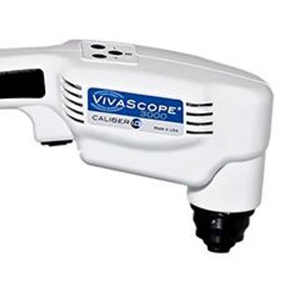
VivaScope - Model 3000 -Specialty Confocal Microscopes
The VivaScope System produces horizontal sections of the skin. Horizontal sections make it straightforward to view the various layers of the skin in sequence, from the outer surface of the stratum corneum, through the granular, spinous and basal layers, to the level of the superficial reticular dermis. The VivaScope System is capable of imaging cells in the epidermis of skin and the fibrous tissue, primarily collagen and fibrin, in the dermis. In addition, it is possible to visualize circulation of blood cells, including both erythrocytes and leukocytes, in capillaries and other small vessels.
Image Format and Resolution
- Imaging Wavelengths: 830nm
- Optical Resolution (in tissue):
Horizontal: <1.25 μm at center of field of view (FOV)
Vertical: <5.0 μm at center of FOV - Single Frame FOV: 750 μm x 750 μm
- Displayed Image Resolution: 1000 x 1000 pixels
- Depth of Imaging: Superficial Reticular Dermis; dependent on tissue
- Image Formats: DICOM-compatible bitmap/DICOM-compatible movie
Environmental
- Operating Environment: 55° to 85° F (13° to 30° C)
- Operating Humidity: Non-Condensing
- Power Source: 110-240V, 50-60Hz
Laser System Classification: CDRH USFDA Class I, IEC Class 1M
Regulatory Certifications and Standards: FDA 510(k) cleared, CE Mark
Physical
- Base Dimensions: 56.89" x 25"
- Handheld
- Also available as an option in combination with the VivaScope 1500
- FDA 510(k) cleared
- Compact microscope design
- Three image capture modes:
- VivaStack®: Stack of multiple images penetrating into the epithelium and supporting stroma
- Confocal Capture: Single 750 μm X 750 μm confocal image capture
- Movies: Live video stream capture