

BESA - Version Research 7.1 -State-of-the-art Research Tools
BESA Research 7.1 provides the ideal combination of state-of-the-art research tools and proven diagnostic workflows in M/EEG analysis of human brain signals. Whether you are aiming for unlocking mechanisms of brain connectivity and function, or for enhanced diagnostic use on epileptiform data, BESA Research 7.1 will help you to achieve your targets reliably and with more pathways than ever before. This further expands on the set of new features that was introduced in the previous version BESA Research 7.0.
Source analysis and source imaging
- For the first time, Boundary element method (BEM) and Finite element method (FEM) models can be directly compared on M/EEG data with just a couple of mouse clicks! After calculating the models automatically in BESA MRI, the models are automatically loaded into the Source analysis module and can be exchanged by two clicks in the head model section of the module.
- MEG and EEG can now be combined into a single data set for simultaneous source modeling of the two modalities. This is available for many source analysis and source imaging methods.
- MRI display now offers multi-slice view in any orientation for an easier review of solutions in the individual anatomy.
- Confidence limits are calculated for discrete source solutions, and displayed in co-registered MRI images.
- The Bayesian source imaging method SESAME was improved to enhance robustness, as well as speed of computation and convergence. For this purpose, hyper-priors were introduced (cf. https://arxiv.org/abs/2006.04141) and parallel computing was optimized for this method.
- The full noise covariance matrix computed from individual trials can now be used in computation of minimum norm estimates.
- Calculation of beamformer virtual sensor montages based on atlas regions is now supported.
- Two new brain atlases were added: Yeo7 and Yeo17 (https://doi.org/10.1152/jn.00338.2011).
- Montreal Neurological Institute (MNI) coordinates can now be used in the Source Analysis window.
- The baseline interval definition now features an automatic alert if it interferes with signal of interest.
- Ready-made color schemes for publication purposes are now available.
Data review and pre-processing
- Atlas-based source montages: Pre-computed atlas-based source montages are now available from the menu entry Montage/Source/Atlas montages as well as under the Src button in the control ribbon.
- Parallel computing is used for speed-up of many time-consuming tasks.
- Smoother and faster plotting of waveforms eases review of high-density M/EEG data.
- New data readers for XDF and Neuroscan CURRY 8 formats are available.
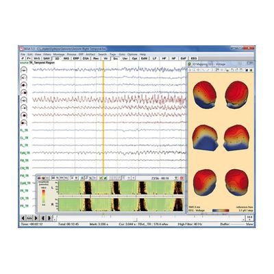
Data review and processing module
BESA Research provides many tools for reviewing and processing of your EEG or MEG data. Raw data are directly read using readers implemented for many EEG and MEG file formats. Data processing steps include digital filtering, artifact detection and correction, computation of correlation and spectral analysis. All these steps can be performed easily with a few mouse clicks. A variety of different display options allows for convenient review of your data.
Data import and export
- Direct readers for most EEG and MEG data file formats
- Import of user-defined file formats using generic reader
- Data import / export to ASCII and binary files
- MATLAB interface for direct transfer of analysis results to MATLAB
Data processing
- Superior digital filtering: high, low, and narrow band pass, notch
- Interpolation from recorded to virtual and source channels
- Automated EOG and EKG artifact detection and correction
- Advanced user-defined instantaneous artifact correction
- Pattern detection and averaging by spatio-temporal correlation
Data review
- Easy and fast review of digital EEG and MEG data files
- Fast paging, tagging and selected viewing of epochs of interest
- DSA and event displays for quick jump to relevant pages
- Additional selected and virtual artifact channels (EOG etc.)
- Linear and non-linear correlation between scalp and source channels
- Spectral analysis: FFT, DSA, power and phase mapping
- Independent Component Analysis (ICA): Decomposition of EEG/MEG data into ICA components that can be used for artifact correction and as spatial sources in the source analysis window