
- Home
- Companies
- Ryan Hennessy Wastewater Microbiology
- Software
- Ryan-Hennessy - Version 1.0 - Machine ...


Ryan-Hennessy - Version 1.0 -Machine Learning Tool
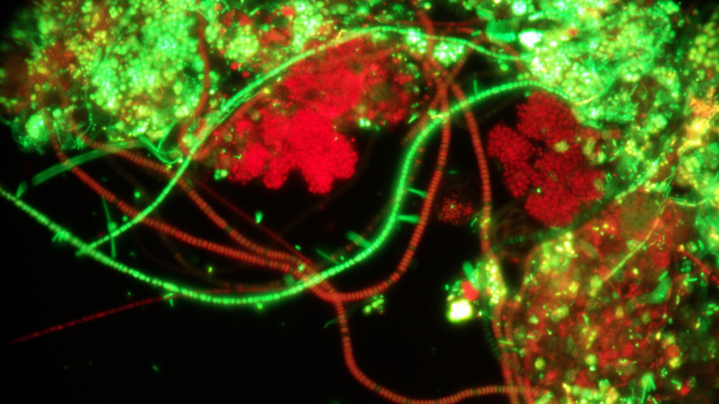
Version 1.0 of this model has been trained using thousands of pictures taken by Ryan Hennessy Wastewater MIcrobiology from a wide range of municipal and industrial wastewater treatment systems throughout predominantly the United States and Canada. The model has been trained for a high variety of microbe types and conditions which include the following list and recommended magnifications for optimal usage:
This is a 1.0 version of the machine learning model and future updates may occur depending upon user interaction rates and feedback, it cannot be guaranteed 100% accuracy. Upon submitting your picture, the AI will generate an email that Ryan Hennessy is also copied on. Ryan will review the email, and in the event that there is disagreement with the model, he will reach out to you at an estimated time of between 10 minutes to 48 hours.
Please note that this machine learning model is designed as an operational tool for recognizing 1 microbe type or condition at a time per picture and is often NOT representative of overall plant conditions. For any microbe type or condition and its associated growth cause (s) to be relevant it must be ranked common or greater in abundance using the Jenkins ranking system. This model is intended for helping professionals with identification of unknown items they view under the scope. For an overall "big picture" diagnosis of treatment plant conditions a full microscopic evaluation by a trained professional is recommended in which items such as floc structure, dispersed growth, higher life form organisms, filament impact on floc structure, polysaccharide, rank/type of filamentous bacteria types and other indicator organism phenotypes, Gram staining, Neisser staining, and (preferably fluorescent viability testing). Information for sending a sample for microscopic evaluation is available at: Services - Ryan Hennessy Wastewater Microbiology
Microbe Type or Condition: Recommended Photo Options
- Actinomycetes_Mycolata (includes Nocardioforms): 1000x phase contrast oil immersion, Gram stain 1000x
- Anaerobic sulfur bacteria: 1000x phase contrast oil immersion
- Anaerobic filament morphotype B: 1000x phase contrast oil immersion
- Beggiatoa: 1000x phase contrast oil immersion or 400x phase contrast
- Bristleworm: 100x or 200x phase contrast or brightfield
- Crustacean: 4x, 100x, or 200x phase contrast or brightfield
- Dead filament: 1000x phase contrast oil immersion
- Dispersed growth high: 100x or 200x phase contrast
- Dispersed growth moderate: 100x or 200x phase contrast
- Dispersed growth low: 100x or 200x phase contrast
- Elevated polysaccharide: 100x, 200x, 400x reverse India ink
- Ferric iron: 100x, 200x, 400x, and 1000x phase contrast
- Fibrous material: 100x or 200x phase contrast, possibly 100x or 200x brightfield (lower probability of success)
- Filament bacteria impact on floc structure high: 100x or 200x phase contrast, possibly 100x or 200x brightfield and possibly 400x phase contrast and 400x brightfield
- Filamentous bacteria impact on floc structure moderate: 100x or 200x phase contrast, possibly 100x or 200x brightfield and possibly 400x phase contrast and 400x brightfield
- Filamentous bacteria impact on floc structure low: 100x or 200x phase contrast, possibly 100x or 200x brightfield and possibly 400x phase contrast and 400x brightfield
- Note: It is highly likely that at lower power magnification (100x, 200x) that the machine learning may recognize multiple conditions and lower % (i.e., dispersed growth and floc structure)
- Flagellate: 100x phase contrast, 100x brightfield, 200x phase contrast, 200x brightfield, 400x phase contrast, 400x brightfield, 1000x phase contrast, 1000x brightfield
- Flexibacter: 1000x phase contrast, possibly 1000x brightfield
- Floc open/diffuse: 100x or 200x phase contrast, possibly 100x or 200x brightfield and possibly 400x phase contrast and 400x brightfield
- Floc strong: 100x or 200x phase contrast, possibly 100x or 200x brightfield and possibly 400x phase contrast and 400x brightfield
- Floc weak: 100x or 200x phase contrast, possibly 100x or 200x brightfield and possibly 400x phase contrast and 400x brightfield
- Free swimming ciliate: 100x, 200x, 400x, and 1000x phase contrast and brightfield
- Fungi: 100x, 200x, 400x, and 1000x phase contrast and likely brightfield
- GAO: 1000x Neisser
- Gastrotrich: 100x, 200x, 400x, and 1000x phase contrast
- Grease: 100x phase contrast cropped, 200x phase contrast 1000x phase contrast
- Haliscomenobacter: 1000x phase contrast
- Hyphomicrobium: 1000x phase contrast
- Inert material: 200x phase contrast possibly 200x brightfield
- Iron sulfide: 100x phase contrast cropped, 200x, 400x, and 1000x phase contrast
- Irregular growth formations: 1000x phase contrast
- Microscrilla: 1000x phase contrast
- Microthrix: Gram stain 1000x, 1000x phase contrast, Neisser 1000x (limited success)
- Normal polysaccharide: 100x, 200x, 400x reverse India ink
- Nostocoida limicola: 1000x phase contrast, moderate accuracy at 1000x Gram stain and 1000x Neisser stain
- Oil: 1000x phase contrast
- Thiopedia: 1000x phase contrast
- Thiothrix: 1000x phase contrast
- Type 021N: 1000x phase contrast, possibly 400x phase contrast and 1000x brightfield
- Type 0041/0675: 1000x phase contrast
- Type 0092: Neisser 1000x
- Type 0581_Chloroflexi: Best at Gram stain 1000x, low likelihood of recognition at 1000x phase contrast
- Type 0914/0803: 1000x phase contrast
- Type 0961: 1000x phase contrast
- Type 1851: 1000x phase contrast
- Type 1863: 1000x phase contrast, 1000x Gram stain
- Water Bear: 100x, 200x, 400x, 1000x phase contrast and brightfield
- Yeast: 400x phase contrast, 1000x phase contrast
- Zoogloea: 100x phase contrast (low success), 200x, 400x, and 1000x phase contrast