
Vuno Med - Version Fundus AI -X-ray Image Analysis Software
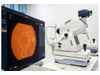
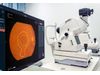
VUNO Med-Fundus AI provides major findings necessary for the diagnosis of retinal diseases based on fundus images.
- AI-based diagnostic supporting solution for the fundus of the eye
- Korea`s first innovative medical device
- MFDS(K-FDA) approved
- CE Certified
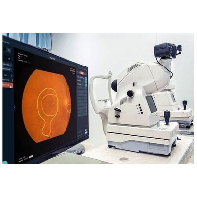
It helps intuitively check the presence of 12 abnormalities and locate them.
- Drusen
- Hemorrhage
- Hard Exudate
- Cotton Wool Patch
- Vascular Abnormality
- Glaucomatous Disc Change
- RNFL Defect
- Membrane
- Chroioretinal Atrophy
- Non-glaucomatous Disc Change
- Macular Hole
- Myelinated Nerve Fiber
The software automatically locates optic discs and macula to mark eight regions of the fundus* and help diagnose the drawn regions.
- Macular
- Superior optic disc
- Inferior optic disc
- Temporal
- Superotemporal
- Inferotemporal
- Superonasal
- Inferonasal
Based on a total of 103,262 fundus images on which 57 ophthalmologists* executed a triple reading, the diagnostic supporting system detects abnormalities in fundus images.
* 16 retina specialists, 9 glaucoma specialists and 3 corneal specialists
References 1. Son J, et al. Development and Validation of Deep Learning Models for Screening Multiple Abnormal Findings in Retinal Fundus Images.Ophthalmology (2019).
VUNO Med-Fundus Al provides a patient report containing visualized abnormality findings that facilitates doctor-patient communication with enhanced patient satisfaction.