

- Home
- Companies
- Edinburgh Instruments - TECHCOMP Group
- Articles
- Map of the Month - June
Map of the Month - June
Welcome to Edinburgh Instruments monthly blog celebrating our work in Raman, Photoluminescence, and Fluorescence Lifetime Imaging. Every month we will highlight our pick for Map of the Month to show how our spectrometers can be used to reveal all the hidden secrets in your samples.
June
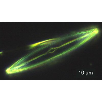
Diatoms are light absorbing algae which, through carbon fixation, produce over 20% of the air we breathe. Their cell walls can be thought of as glass houses, made of silica, they protect the cell inside and produce intricate and striking patterns, Figure 1. Diatoms are an extremely diverse organism; new species continue to be discovered and there is still so much to be learned by scientists about these important algae. They are indicative of water quality due to individual species having distinct ranges of pH and salinity for them to grow, their growth is also dependent on species tolerance levels to other environmental conditions such as nutrient concentration.
Given their importance both in and out of the water they live in, studying diatoms is critical for understanding their roles in the ecosystem. The gold standard methods to assess water quality using diatoms as a bioindicator are time-consuming, expensive, and complex, in contrast Raman spectroscopy is rapid, easy-to-use, and cost effective. When using a confocal Raman microscope, the components of the silica glass wall of an individual diatom can be imaged in 3D, Figure 2, helpful for assessing its thickness whilst obtaining information on the structure.
Edinburgh Instruments Confocal Raman Microscopes
If you are interested in microscopy in forensic science, find out more about our RM5 and RMS1000 Raman Microscopes. If you would like to talk to one of our sales team and find out how our Raman Microscopes can be used within your own research, please contact us.