- Home
- Companies
- Rapp OptoElectronic
- Products
Rapp OptoElectronic products
Microscopy and Imaging - Multi-Photon Microscopy
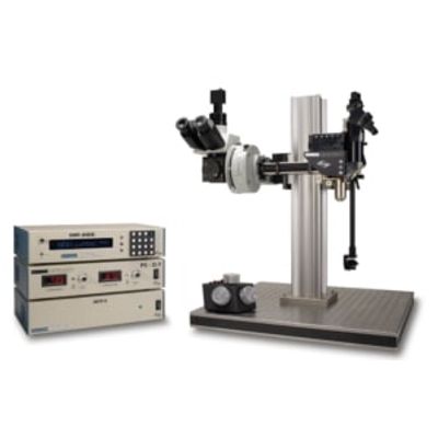
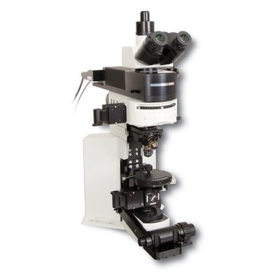
RAPP - Model MOM - Movable Objective Microscope for Two-Photon Imaging
The Movable Objective Microscope (MOM) by Sutter Instrument is a sophisticated two-photon microscope designed for advanced imaging within living specimens. This system features a unique design allowing three-dimensional movement and rotation of objectives while keeping the specimen stationary, optimizing the imaging process. The MOM integrates two independent microscopes: a wide-field section equipped with an Olympus vertical illuminator for standard epifluorescence, and a two-photon section that guides excitation laser light for deep tissue imaging. It supports precise and smooth X, Y, and Z movements with minimal drift and high reproducibility, akin to Sutter's MP-285 micromanipulator. This allows efficient Z-stack and mosaic image acquisition without moving the stage. The flexible horizontal light path facilitates conversion between upright and inverted configurations, suitable for imaging various non-horizontal surfaces. The MOM remains adaptable to evolving scanning technologies, supporting multiple scanner types, and offers customizable detector paths for optimal photon capture. Compatible with ScanImage software, it ensures seamless integration with imaging hardware.