WITec - Model alpha300 apyron -Fully Automated Raman Imaging System
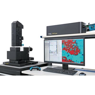
The alpha300 apyron is the top of the line Raman imaging system in WITec’s microscope series. It combines ease-of-use and ultimate capability by automating hardware control and offering pre-configured measurement routines. This streamlines the experimental workflow and yields reproducible results with unrivaled speed, sensitivity and resolution. With the release of a new generation of alpha300 apyron microscopes, WITec now takes Raman imaging automation to the next level.
Since its first release in 2015, the alpha300 apyron has been WITec’s high-end fully-automated Raman microscope. WITec’s apyron systems are always equipped with the most recent features and developments, especially with respect to automation and user comfort.
Extensive automation
- simplifies operation of the instrument.
- requires less human input.
- reduces potential sources of error.
- enhances reproducibility.
- allows for completely remote-controlled use in enclosed environments (i.e. in a glove box).
The alpha300 apyron is the ideal Raman microscope for:
- multi-user laboratories with varying requirements and levels of user experience.
- industry facilities with recurring experimental situations and an emphasis on time-critical turnover.
- Raman newcomers with advanced imaging requirements.
- veteran Raman microscopists seeking the next performance benchmark.
- researchers employing remote operation, such as in enclosed environments.
In the alpha300 apyron, the entire workflow from system calibration through Raman measurements to data analysis is fully automated and can be completely remote-controlled. You never have to touch the microscope – except to change your sample.
Setup
- TrueCal executes pre-configured calibration routines.
- Laser safety class 1 (or 1M) compliant.
- AutoBeam concept for automatic and motorized beam path alignment includes:
- Motorized 6-position objective turret: positions selected objective and compensates offsets
- Motorized illumination selector: switches between all microscopy illumination options
- Motorized laser coupler: delivers up to 6 laser wavelengths from UV to NIR
- AutoBeam output coupler: automatically optimizes signal & resolution and selects spectrometers
- Motorized calibration source: fast & automatic multi-point spectrometer calibration over the full spectral range
- Motorized Köhler illumination apertures: facilitate focusing and optimize contrast and homogeneity
- Motorized polarization modules: freely rotatable automated excitation polarizer and detection analyzer
- TruePower sets and determines absolute laser power with
- Instrument (re-)positions sample automatically with motorized and piezo-driven scan stages.
- Focus stabilization finds and actively maintains focus during the entire measurement.
- Motorized objective turret includes precise offset-compensation – AFM/SNOM objective-compatible.
- EasyLink handheld controller offers intuitive instrument operation.
Data processing and display
- Suite FIVE software wizard guides the user through the processing of Raman spectra, from background reduction through image generation.
- TrueComponent Analysis: automatically locates the sample components in an image and differentiates their individual spectra.
- TrueMatch software identifies molecules by comparing measured spectra with existing databases.
The alpha300 apyron sets the benchmark for automated Raman imaging systems with unrivaled imaging qualities. It simultaneously offers the highest spectral and spatial resolution, measurement speed and signal sensitivity. All parts of the system are optimized for the highest transmission efficiency and precision. Additionally, the modular design ensures freedom in system configuration to fulfill the specific requirements of your applications, even as they evolve. A wide variety of excitation lasers, ultra-high throughput spectrometers (UHTS) and detector types are available.
True confocality and wavelength-optimized design
- guarantees maximum throughput.
- provides sharp 2D and 3D images with resolution limited only by physical law:
- lateral resolution
- depth resolution
- yields spectral resolution down to 0.1 cm-1 relative wavenumbers (at 633 nm excitation).
- supports ultra-fast Raman imaging without sacrificing resolution (acquisition speed
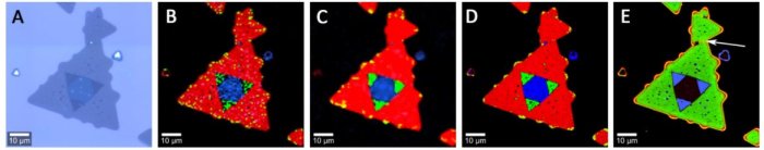
The extremely high performance and speed of the alpha300 apyron microscope is vividly demonstrated with the analysis of a tungsten diselenide (WSe2) flake. The flake’s different layers are visible in the white-light image (A) and can be characterized in more detail by Raman imaging. In only about 2 minutes, a clear and informative 75 x 75 µm² Raman image consisting of 10,000 spectra was recorded (B). The flake consists of single-layer (red), double-layer (green) and multi-layer (blue) areas. The same measurement after smoothing is shown in (C). A measurement of about 17 minutes consisted of more than 100,000 spectra and produced an even sharper image (D). The increased signal to noise ratio was achieved by reducing the pixel size from 750 nm (B) to 230 nm (D). The photoluminescence image (E) shows the same structures as the Raman image and even the grain boundary between the large and the smaller flake is visible. The integration time was 6 milliseconds per pixel in all measurements.
Carbon tetrachloride (CCl4) is a suitable reference sample for determining the performance of a Raman spectrometer in terms of spectral resolution. The characteristic peak at 460 cm-1 should be clearly resolved into three peaks at room temperature. Due to its ultra-high optical throughput the alpha300 apyron allows for ultra-fast Raman imaging at high spatial resolution while simultaneously maintaining the ability to resolve this spectrum.
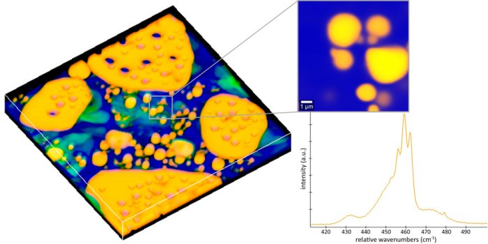
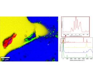
Raman image of a solution of L-cysteine in a mixture of water and different herbal oils. Most of the L-cysteine is dissolved in the water phase (blue). In the oil phase (yellow), an undissolved cysteine crystal (red) is visible. The green areas likely represent one of the herbal oil ingredients. The Raman spectrum of L-cysteine has characteristic peaks in the low-frequency range, which were clearly resolved by the alpha300 apyron equipped with the WITec RayShield coupler.
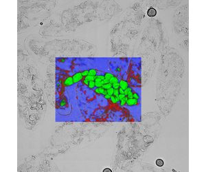
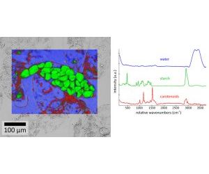
Overlaid white-light and confocal Raman images of squashed banana pulp. This high-resolution Raman image consists of more than a million spectra and was acquired in only 36 minutes. Carotenoids (red), starch (green) and water (blue) were the most prominent ingredients.
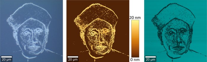
White-light image (left), AFM image (middle) and Raman image (right) of a portrait of C. V. Raman on a silicon chip. The image is part of a calibration standard for Raman microscopes, produced and provided courtesy of the Physikalisch-Technische Bundesanstalt (PTB) in Braunschweig, Germany.
WITec’s fully-automated alpha300 apyron Raman imaging system won the first prize at the Achema 2015 Innovation Awards in the category of Laboratory and Analytical Technologies. Achema, in Frankfurt (Germany), is the world’s largest trade show for the chemical engineering and process industry. The Innovation Awards are bestowed by the renowned industry experts and technical editors of: Process, Process Worldwide, PharmaTec, Process China, Process India and Laborpraxis.