

- Home
- Companies
- IXRF Systems Inc
- Products
- Model SEM-XRF - X-ray Sources for ...
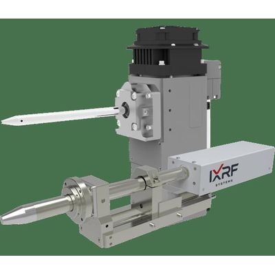
Model SEM-XRF -X-ray Sources for Electron Microscopy (SEM)
microXRF X-ray sources for Scanning Electron Microscopes (SEM): addition of an polycapillary X-ray tube and Iridium Ultra software will transform your SEM’s quantitative analytical capabilities. Higher peak to background ratios enable greater elemental sensitivity for higher Z elements: sensitivity exceeding e–-beam excitation by a factor of 10-1000X. Exceptional beam stability, together with a modern SDD X-ray detector, afford higher precision with ppm-level sensitivity. Non-conductive materials may be analyzed without any special preparation or coating. We integrate with your SEM to deliver full spectrum analysis using excitation from both the e–-beam and our X-ray source.
Distribution analysis stores complete spectra for every map point for on- and off-line analysis. Samples can be analyzed with micro-spot X-ray beam and e?-beam simultaneously without position change. Both excitation methods are integrated in our analytical software suite. No interference with normal SEM operation, the X-ray source can stay in its measurement position permanently. microXRF is completely non-destructive and does not require the sample to be coated. Use your existing EDS detector system; we support most all microscopes.
- Analytical results compare to those of standalone μXRF systems
- State-of-the-art analytical software: Iridium Ultra
- Decades of continuous development & innovation
- Selectable primary X-ray filters
- To suppress Bremßtrahlung and diffraction peaks
- For lower detection limits, down to PPM levels for most elements
- Uses your scanning electron microscope’s motorized stage
- Allows sample tilt to produce minimum spot sizes
- No special cooling is required, our sources are air cooled
Another advantage of having micro-XRF in a scanning electron microscope (SEM) is the ability to employ the characteristic lines from the anode material (typically Ag, Cu, Mo, Rh or W) of a X-ray tube to non-destructivity determine the average atomic number of any phase or artifact. The high specificity of elastic (Rayleigh), as well as inelastic (Compton), X-ray scattering to the mean atomic number of a specimen has long been recognized.1 Micro-spot X-ray fluorescence (micro-XRF) may thus be exploited to non-destructively gain more information on chemical composition. In practice, the Rayleigh/Compton intensity ratio from XRF spectra may be calibrated, with standard reference materials, relative to mean atomic number. This sort of calibration curve can reveal valuable information on the elemental composition complementary to that obtained from conventional elemental analysis via XRF-SEM and/or SEM/EDS. Particularly for matrices of lower mean atomic numbers (like plastics), the sensitivity of the approach is so high that it can be easily distinguish between specimens of mean atomic numbers differing from each other by 0.1 Z. Hence, the content of light elements which are “invisible” for XRF, particularly hydrogen, or of heavier impurities/additives in light materials can be calculated “by difference” from the scattering calibration curve.