

- Home
- Companies
- Rapp OptoElectronic
- Products
- RAPP - Model DF-Scope - Multi-Photon ...
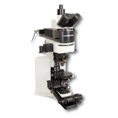
RAPP - Model DF-Scope -Multi-Photon Imaging Package
The DF-Scope is a specialized multi-photon imaging package developed for the Olympus BX51WI upright microscope, designed specifically for laboratories engaged in electrophysiology and epifluorescent imaging. This package enhances the microscope's capabilities by integrating components essential for multi-photon imaging, leveraging a Ti:Sapphire laser for optimal performance. Key features include the incorporation of elements from the MOM system such as resonant and galvo scan boxes, controllers, detector paths, and high-sensitivity photomultiplier tubes (PMTs). These components facilitate the capture of light emitted from the sample's focal volume, a critical aspect of non-linear excitation imaging. The DF-Scope is equipped with dual detector paths, one positioned above and one below the sample, to maximize light collection. This configuration is especially beneficial for thin samples, such as brain slices, by utilizing sub-stage detectors for enhanced signal acquisition, adaptable with various Olympus condensers.The DF-Scope™ is a customer-inspired, multi-photon, imaging package for the ubiquitous BX51WI upright microscope. Many laboratories already use BX51WI microscopes for electrophysiology and epifluorescent imaging experiments. The DF-Scope package provides the necessary optics and electronics for the BX51WI to be used for multi-photon imaging (with the addition of a Ti:Sapphire laser). The design incorporates subassemblies from our MOM™ (Movable Objective Microscope®) system including resonant and galvo scan boxes and controllers, detector paths, PMTs, PMT power supplies, scan lenses and tube lenses.
A hallmark of multi-photon imaging is that all of the light emitted by the sample is known to emit from the focal volume as a result of the non-linear excitation of the fluorophore. High sensitivity photomultiplier tubes (PMTs) are used to collect as many photons as possible in order to reconstruct the scanned image. The DF-Scope design allows for two detector paths to gather more emitted light – one above the sample and one below. If using a thin sample, like a brain slice, we recommend the lower (sub-stage) detectors for increased signal detection. Additional signal will be available at the trans detector path. This substage detector assembly is designed to work with a variety of Olympus condensers.
NOTE: The DF-Scope design requires an Olympus BX51WI and the following Olympus parts: WI-ARMAD, 5-UR710LP and U-M619.
- Converts a standard Olympus BX51WI into a two-photon microscope while retaining standard microscope functions (transmitted light and epifluorescent imaging).
- Includes detector(s) and “whisperquiet” resonant scan box developed for the MOM™ Two-Photon microscope
- Upper and lower photodetectors for increased collection efficiency
- Fully compatible with the Sutter MPC-78 Large Moving Stage Platform and motorized focus drive
- Designed to be controlled with the Sutter MCS (MOM Computer System) Imaging Software including resonant scanning with MScan 2.0
- Also fully compatible with most multi-photon freeware such as ScanImage 5.0, Helioscan, and MPScope
- Breadboard format in scan pathway allows easy addition of photostimulation light sources to the main scanned laser path
- In vivo and in vitro two-photon imaging
- Whole-cell imaging
- Intracellular imaging