- Home
- Companies
- Creative Proteomics
- Services
- Sample Preparation Services
Sample Preparation Services
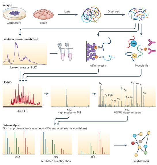
Sample preparation is critical for proteomics analysis, and Creative Proteomics provides sample preparation service according to your needs.Proteomics aims to study dynamically changing proteins expressed by a whole organism, specific tissue or cellular compartment under certain conditions. Consequently, two main goals of proteomics research are to identify proteins derived from complex mixtures extracted from cells, and to quantify expression levels of those identified proteins.
Each of these protein sources is comprised of many more components than the proteins themselves, these non-protein sources include nucleic acids, lipids, sugars, and a wide variety of small molecules. Furthermore, unlike DNA and RNA, both of which are chemically homogeneous, proteins are very heterogeneous and vary widely with respect to their size, charge, hydrophobicity, and structure. In addition, many proteins undergo post-translational modification and may have a variety of non-peptide moieties bound to them. This chemical heterogeneity makes isolating proteins extremely challenging.
In proteomics analysis, sample preparation is the most sensitive step in the entire procedure, and here are a few tips. The treatment of the sample must be kept to a minimum to avoid protein losses; the sample should be kept as cold as possible to avoid protein modifications and prevent enzyme activities; the preparation time should be kept as short as possible to avoid losses and modifications; all aggregates and complexes should be disrupted; disturbing compounds should be removed without removing proteins. It should also be noted that there are more than one possible procedure to treat a sample, and a 100% representation of the proteins contained in a cell will never be obtained in practice. Usually the method, which will display the highest number of different proteins, is chosen.
Proteome is the collection of gene products expressed at both low levels and high levels, especially, the reliable and quantitative comparison of low-abundant proteins is a tremendous challenge of proteomic analysis, thus the sample preparation techniques of greatest interest in expression proteomics focus on pre-fractionating and enriching proteins before their separation. Highly abundant proteins such as actin and tubulin or human serum albumin, proteins of the complement system and antibodies will comprise a significant portion of this homogenate, and the less-abundant regulatory proteins that are most informative regarding cellular response can be detected more efficiently with proper pretreatment and handling.
Subcellular fractionation can be conducted as a means to enrich specific organelles and fractions, and thus enable the visualization of significantly more proteins than can be detected in a whole homogenate. Different subcellular compartments contain not only different but also compartment-specific subsets of gene products in order to provide suitable biochemical environments, in which they exert their particular function. The identification of subsets of proteins at the subcellular level therefore represents an initial step towards the understanding of cellular function. Indeed, there are subsets of proteins that are associated with subcellular structures only in certain physiological states, but localized elsewhere in the cell in other states.
The proteins have to be extracted from cells or tissue material and the choice of disruption methods is dependent on the type of sample. The easiest way is osmotic lysis of cultured cells using the standard lysis solution, while tissues and samples with tough cell walls are often treated with mortar and pestle at sub-zero temperatures.
Unfortunately it is impossible to separate complex protein mixtures under native conditions. Liquid samples have to be denatured to prevent the formation of polypeptide oligomers, aggregates and interactions. In presence of more than 7M urea, most polypeptides exist only in one single configuration. Also the addition of carrier ampholytes supports the solubility of hydrophobic proteins. The reductants DTT or DTE prevent different oxidation steps of the proteins. Some proteases are also active in presence of urea and detergents, protease inhibitors can inactivate most of the proteolytic activities, in practice the sample is extracted with a protease inhibitor cocktail, and then the sample is precipitated.
A crude extract can be contaminated with salt ions, phospholipids and nucleic acids, leading to disturbances or background streaking. Nucleic acids are visible after silver staining as disturbing horizontal streaks in the acidic part of the gel. Furthermore, they can precipitate with the proteins when the sample has to be applied on the basic end of the IEF gel. They can be removed with DNAseI and RNAseA treatment, or with benzonase. The easiest technique is sonication, which breaks nucleic acids into little fragments. Precipitation also removes nucleic acids. Lipids are removed with an excess of detergent (>2%) or with precipitation, proteases are irreversibly inactivated by precipitation, solid material is removed by spinning it down by centrifugation, and salts can be dialyzed away or removed by precipitation.