

- Home
- Companies
- AAT Bioquest, Inc.
- Products
AAT Bioquest, Inc. products
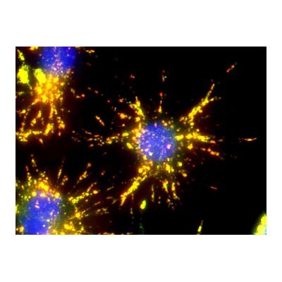
Bioconjugation - Biotin and Streptavidin
AAT Bioquest - Biotin-Streptavidin Conjugation System
The widespread adoption of the biotin-streptavidin conjugation system is primarily due to two factors. The first is the relatively small size of the biotin and streptavidin molecules themselves, which allows for extensive binding to biologically active macromolecules, such as antibodies, without impedance to their functions (i.e. an antibody`s binding site). The second is the specificity with which biotin and streptavidin bind to each other, as well as the strength of the subsequent bond, which allows for powerful, stream-lined bioassay applications. Streptavidin tetramers have an extraordinarily high binding affinity for biotin with a dissociation constant (Kd) of approximately ~10-14 mol/L. This tight and specific binding is rapid and able to withstand extremes in pH, temperature, organic solvents, and denaturing reagents.
Physiological Probes - Calcium Indicators
AAT Bioquest - Quantifying Cytosolic Calcium
Calcium is a ubiquitous messenger impacting a number of signal transduction pathways including calcium-induced changes in protein confirmation, the regulation of cytosolic and organelle calcium levels, the highly localized nature of calcium-mediated signal transduction and its specific roles in excitability, exocytosis, motility, apoptosis, and transcription. These pathways regulate many key cellular processes including neurotransmission, cell proliferation, and muscle contraction. Under normal conditions with respect to the extracellular fluid, the resting cytoplasmic concentration of calcium is relatively low, at or below 100 nM. Calcium signaling is activated either through the release of calcium ions from intracellular stores, or by an influx of extracellular calcium through plasma membrane ion channels. Stimulated cells can increase cytoplasmic calcium concentration up to 500 to 1000 nM.
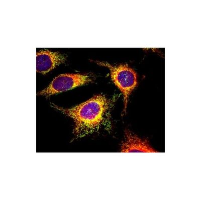
Bioconjugation - iFluor™ Dyes and Kits

AAT iFluor - Reactive Dyes
iFluor™ reactive dyes may be covalently labeled to biomolecules without self-quenching, producing intensely fluorescent conjugates. They are widely used to modify amino acids, peptides, proteins (in particular antibodies), oligonucleotides, nucleic acids, carbohydrates and other biological molecules. iFluor™ reactive dye formats include amine-reactive succinimidyl ester, thiol-reactive maleimide, and more.
Antibodies and Proteomics
Alkaline Phosphatase (ALP)
Antibody and Protein Labeling
ReadiLink - Rapid Antibody Labeling Kits
AAT Bioquest ReadiLink™ Rapid Antibody Labeling Kits provide a quick and convenient method to label microscale volumes of antibodies with our superior iFluor®dyes, mFluor™ dyes, or other labels. The unique chemistry of ReadiLink™ kits enables researchers to effortlessly label and recover 100% of their antibodies without a purification step. Since ReadiLink™ conjugates are covalently labeled, they are stable for long-term storage. They are ideal for demanding applications, including fluorescent microscopy, multi-color imaging, multiplex flow cytometry, primary detection, IHC, FISH, ELISAs, and more.
Chemical Reagents
Model Classic - Dyes
Fluorescence is the result of a three-stage process that occurs in certain molecules (generally polyaromatic hydrocarbons or heterocycles) called fluorophores or fluorescent dyes. Fluorescent probes enable researchers to detect particular components of complex biomolecular assemblies (including live cells) with exquisite sensitivity and selectivity. Reactive fluorescent dyes are widely used to modify amino acids, peptides, proteins (in particular, antibodies), oligonucleotides, nucleic acids, carbohydrates and other biological molecules.
Peptide and Oligonucleotide Labeling
Dye-labeled peptides and oligonucleotides are important tools in biochemical and cellular studies. Fluorescent peptides and oligonucleotides have been extensively used in all major types of fluorescence imaging including fluorescence resonance energy transfer (FRET). These labeled biomolecules are widely used for diagnosing infectious diseases based on the molecular beacon and other technologies. FRET peptides and oligonucleotides have been also used for cell analysis via fluorescence-associated cell sorting (FACS) either in vivo or in vitro for research and diagnostic purposes.
Organelles
ER Tracer - Endoplasmic Reticulum Staining Kits for Live Cells
ER Tracer™ dyes are membrane-permeant stains considerably selective for the endoplasmic reticulum in living cells. As the key component in our Cell Navigator® Live Cell Endoplasmic Reticulum Staining Kits, ER Tracer™ dyes can be multiplexed with other fluorescent proteins or probes in live cell multiparametric studies or after fixation for colocalization studies. However, for certain cell lines, ER Tracer™ stains may not selectively bind to ER.
Molecular Biology
Polymerase Chain Reaction (PCR)
PCR (polymerase chain reaction) is a core technique used extensively in molecular biology research to amplify a specific DNA template in vitro rapidly. It enables researchers to generate significant quantities of sample DNA for a wide range of downstream laboratory and clinical applications, including cloning, genotyping, sequencing, mutagenesis, forensics, and the detection of pathogens to diagnose infectious diseases. Since being introduced in 1985, several iterations of the PCR process have been developed, including quantitative PCR (qPCR) for monitoring DNA amplification in real-time and reverse-transcription PCR (RT-PCR) for the detection of RNA, a tool that has become instrumental in viral diagnostics.